
Nathalie Rademacher, Dr.med.vet., DACVR, DECVDI
Colin Mitchell, BVM&S, DACVS Neely Heidorn, PhD![]()
Original Publish Date December 2014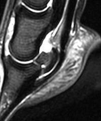
A lameness localized to the hoof based on lameness examination and nerve blocks provides a diagnostic challenge due to the complex soft tissue structures encapsulated within the hoof. Radiography is often the first step for evaluation because it is easy to perform, widely available and can be performed with the horse standing. Radiographs are best for evaluation of bony structures, however subtle changes can require weeks to months before radiographic changes are observed. Contrast radiography can be helpful in cases of penetrating wounds such as stepping on a nail to depict a communication with a joint or tendon sheath. Computed tomography uses the same basic principle as radiography (ionizing radiation), but requires the horse to be anesthetized. This is more sensitive in detection of subtle bony changes, but is limited in the differentiation of soft tissue structures such as tendons, ligaments and joints. Ultrasonography is widely used to evaluate tendons, ligaments and soft tissues but cannot penetrate the hoof capsule.
MRI is a technology that uses a strong magnetic field and radiofrequency waves to produce detailed images that can be acquired in different planes. MRI is very sensitive to changes in the amount of water in tissues and is therefore the method of choice for evaluation of the hoof and its soft tissue structures. Different sequences (a sequence is a set of images) can be acquired that allow tissue characterization and outline disease processes. Once a lameness has been localized to a particular region, an MRI can be performed to investigate cases where a diagnosis cannot be determined by other imaging modalities or for evaluation of a complex lesion. The horses are examined whilst under general anesthesia, in lateral recumbency and average time required for the scan is about 1 hour.
Case Example
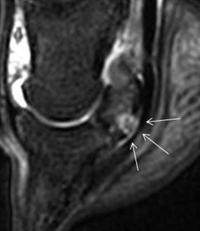
A 3 year old Thoroughbred colt presented with a persistent left front limb lameness. Eight weeks earlier, the horse stepped on a nail and was treated. The perforation of the sole healed without complications but a mild, performance limiting, lameness persisted. MRI of the left front foot was performed as other imaging modalities had failed to identify the cause of this lameness.
See the sagittal image of the distal hoof. A normal horse is shown on in figure 1, and the 3 year old colt’s leg is shown on figure 2. In figure 2, a hyperintense (white) signal within the navicular bone, is marked with the arrows, where the nail caused penetrated the navicular bone.Since bone doesn’t usually contain a lot of water, the typical MRI signal of bone is dark (hypointense). A transverse image of the hoof (not shown), allows you to visualize a focal lesion (sagittal tear in deep digital flexor tendon) within the deep digital flexor tendon that resulted from the previous trauma (stepping on the nail).
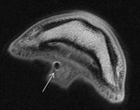
The tract caused by the nail could easily be visualized with the MRI and penetrated the frog (Figure 3, arrow), through the deep digital flexor tendon into the navicular bone. These MRI findings confirmed that a complex injury was present in this horse’s leg. He had an injury involving his navicular bone, the deep digital flexor tendon and the adjacent tissues of his hoof capsule. Without an MRI examination, identifying these lesions would have not been possible. Arthroscopic surgery was performed on the navicular bursa, which allowed the tendon and bone injury to be debrided and an adhesion to be broken down. This procedure allowed the horse to recover from his injury, and with time to return to racing. To schedule an appointment to diagnose a lameness in your horse, please contact: Equine Health Studies Program School of Veterinary Medicine Louisiana State University Baton Rouge, LA 70803, Telephone: (225)-578-9500